Computer Vision-Based Downgaze Palsy Assessment

From manual ruler measurements to a mobile vision pipeline for Progressive Supranuclear Palsy

1University of Richmond 2University of Chicago

Teaser video coming soon

A ruler-based measurement that takes more than five minutes becomes an interactive mobile assessment in under one minute. We then take the next step: automatically detecting the anatomical landmarks needed to estimate downgaze palsy severity from an eye image.

Abstract

Progressive Supranuclear Palsy (PSP) is a rare neurological condition associated with problems in movement, balance, vision, speech, and swallowing. One of its characteristic visual symptoms is downgaze palsy, the reduced ability to move the eyes downward. A clinical image-based assessment can quantify the severity of downgaze palsy using the relative geometry of the pupil, iris, and eye corners, but the existing workflow requires manually overlaying a grid, locating landmarks, measuring distances with a ruler, and computing the final score. We first built an interactive mobile application that digitizes this workflow and reduces the assessment time from more than five minutes to under one minute. We then developed an anatomy-guided computer vision pipeline that automatically detects the pupil, eye corners, and iris top edge, moving toward an automatic assessment that could run in seconds after further refinement and validation.

Privacy notice: Demonstration videos and figures from our interactive application and computer vision pipeline are not shown on this public webpage to protect the privacy of the individuals whose images appear in them.

Can a clinical measurement become a phone-based vision task?

The key measurement is simple in principle, but tedious in practice. Clinicians first identify the external and internal eye corners, the pupil center, and the top edge of the iris. They then construct a horizontal eye-corner baseline and a perpendicular line through the pupil center. The vertical distance from the baseline intersection to the iris top edge is normalized by the iris radius to produce a scale-independent severity measure.
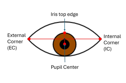
Geometry used to measure downgaze palsy severity

Geometry of the downgaze palsy severity measurement.

Let $p_{EC}$ and $p_{IC}$ denote the external and internal eye corners, $c$ the pupil center, and $t$ the detected iris top edge.

$$\ell(s)=p_{EC}+s\left(p_{IC}-p_{EC}\right)$$

The line $\ell$ defines the eye-corner baseline. Let $\hat n$ be its unit normal, and let the normal through the pupil center be

$$n(\lambda)=c+\lambda\hat n.$$

If $q=\ell\cap n$, the geometric displacement is $a=\|t-q\|$. With iris radius $r$, the severity measure used in the project is

$$\boxed{\text{Downgaze Palsy Severity}=\frac{a}{r}\times 100\%}.$$

From manual measurement to automatic vision

The original ruler-and-grid workflow is slow and sensitive to how the ruler and image are aligned. We therefore developed a classical computer vision pipeline to detect the required anatomical landmarks automatically and use the same clinical geometry to calculate severity.

The first interactive application

Instead of superimposing a grid and measuring the image by hand, the application guides the user through the same anatomical measurement digitally. The user captures an image, selects the required landmarks, and receives the computed downgaze palsy severity immediately. This creates a consistent digital workflow while preserving the underlying clinical geometry.

01

Capture
Take an image of the eye.

02

Select landmarks
Mark the anatomical points required by the measurement.

03

Get the result
Compute the severity measure directly in the application.

Toward an assessment in seconds

> 5 min

Manual grid + ruler workflow

< 1 min

Interactive mobile application

< 5 s

Target once the CV pipeline is integrated into the mobile application

The first timing improvement is demonstrated by the current interactive application. Once the CV pipeline is refined, validated, and integrated into the mobile application, the complete workflow is expected to move directly from image capture to automatic landmark detection and severity estimation in under five seconds.

Automating anatomical landmark detection

Our computer vision pipeline decomposes the problem into three anatomy-guided stages: pupil detection, eye-corner detection, and iris top-edge detection. Rather than estimating the severity score directly from pixels, the pipeline explicitly recovers the same anatomical landmarks used by the manual measurement.

Pupil

Thresholding, contour extraction, contour refinement, ellipse fitting

Corners

Edge preprocessing, Harris candidates, clustering, geometry + intensity filtering

Iris edge

Search for the iris-to-sclera intensity transition along the pupil normal

1. Find the pupil

The eye image is converted to grayscale and binarized to isolate the dark pupil region. We find the largest relevant contour, refine its contour points, and fit an ellipse to recover the pupil center and radius used by the downstream geometry.

2. Find the eye corners

Eye-corner detection begins with grayscale conversion, denoising, contrast enhancement, binarization, edge extraction, and anatomical masking. Harris corner detection supplies candidate points. We cluster those candidates, reject geometrically implausible groups, and use local intensity information to select the final external and internal eye corners.

3. Find the iris top edge

Once the eye-corner baseline and pupil center are known, the final landmark is constrained to a one-dimensional anatomical search. We construct the normal to the eye-corner baseline through the pupil center and search along this line for the intensity transition from iris to sclera. That transition gives the iris top edge required by the severity equation.

The vision model mirrors the measurement itself

A useful property of this pipeline is interpretability. Every stage corresponds to a visible anatomical landmark, and the final score is produced by the same geometry used in the manual assessment. If the output is wrong, we can inspect whether the failure came from the pupil, a corner, the iris edge, or the final measurement. This is especially valuable for a clinical research prototype where transparent failure analysis matters.

Results and validation

The automatic computer vision pipeline has been implemented for the required landmarks and tested on clinical images of patients affected by downgaze palsy. Once the CV pipeline is refined, validated, and integrated into the mobile application, it will automate landmark detection and severity estimation within the existing mobile workflow. Future evaluation will quantify landmark localization accuracy, severity-score agreement with expert measurements, runtime on-device, robustness to image quality and lighting, and end-to-end usability.

Toward assessment in seconds

The next step is to refine and integrate the computer vision pipeline into the mobile application so that it can proceed directly from image capture to landmark detection and severity estimation. With validation, this could make the assessment substantially faster and easier to reproduce. The current system remains a research prototype.